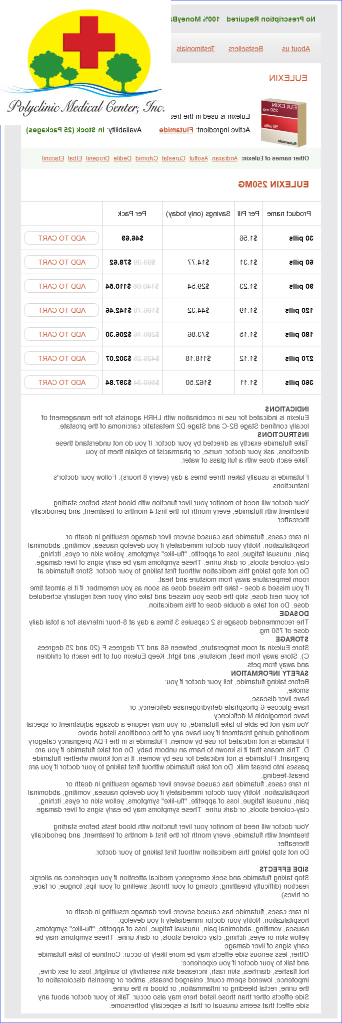
Eulexin dosages: 250 mg
Eulexin packs: 30 pills, 60 pills, 90 pills, 120 pills, 180 pills, 270 pills, 360 pills
Only $1.17 per item
In stock: 706
Description
Within the visual cortex androgen hormone tablets eulexin 250 mg buy with mastercard, the macula of the retina is represented in the posterior half and the paramacular and peripheral parts of the retina are represented successively more anteriorly. The visual pathway includes two parallel streams of information, one concerned with localizing where objects are in the visual field and the other concerned with identifying what the objects are. The "where" stream is the magnocellular (M) path that arises from larger retinal ganglion cells that project to the magnocellular layers of the lateral geniculate nucleus. The "what" stream is the parvicellular (P) path that arises from smaller retinal ganglion cells that project to the parvicellular layers of the lateral geniculate nucleus. Both streams are intermingled in the optic radiation, but each terminates in separate layers of the primary visual cortex. From here, the paths pass to the lateral surface of the cerebral hemisphere, the magnocellular path dorsally to the posterior parietal lobe, and the parvicellular path ventrally to the posterior part of the temporal lobe. Chapter 14 the Visual System: Anopsia Left Right Upper Visual field Lower Left Right Peripheral Paramacular Macular 187 Lens Visual field projection on left retina Visual field projection on right retina Optic tract Loop of Meyer Lateral geniculate nucleus Optic radiation, ventral part (in termporal lobe) Optic radiation, dorsal part (in parietal lobe) A. Knowledge of the representation of the fields of vision in the visual paths is of medical importance. The field of vision is divided into four quadrants: upper right, upper left, lower right, and lower left. The quadrants are demarcated by imaginary horizontal and vertical lines through the fixation point, that is, the point on which vision is focused. Within the optic chiasm, the optic nerve fibers from the nasal or medial halves of the retinae cross, but those from the temporal or lateral halves of the retinae do not cross. This partial decussation serves to bring all of the optic nerve fibers transmitting impulses from either the right or the left half of the field of vision into the contralateral optic tract. Moreover, due to the point-to-point relations that exist between the retina, lateral geniculate nucleus, and primary visual cortex, impulses from the upper and lower halves of the visual field are located in different parts of the optic radiation. Impulses from the contralateral upper quadrant take a ventral course and sweep into the white matter of the temporal lobe before proceeding posteriorly into the occipital lobe where they end in the lower wall of the calcarine sulcus, the lingual gyrus. Impulses from the contralateral lower quadrant, however, take a dorsal course and sweep posteriorly through the white matter of the parietal lobe to the occipital lobe, where they end in the upper wall of the calcarine sulcus, the cuneus. Visual defects are homonymous when confined to the same part of the visual field in each eye. They are heteronymous when the part of the visual field lost in each eye is different. A homonymous defect results from lesions in the visual pathway distal to the optic chiasm. Thus, total destruction of the optic tract, lateral geniculate nucleus, geniculocalcarine tract, or visual cortex results in loss of the entire opposite field of vision in each eye, a phenomenon referred to as contralateral homonymous hemianopsia. Most commonly, the crossing fibers are involved, and this results in an interruption of the nasal retinal fibers, which are carrying impulses from the temporal fields of vision.
Syndromes
- Inability to hold in feces (fecal incontinence) or urine (urinary incontinence)
- Headache
- Weight loss
- Death
- Weakness
- Washing of the skin (irrigation) -- perhaps every few hours for several days
- Coma
- Wheezing
- In the past, most patients with heart valve problems such as mitral stenosis were given antibiotics before dental work or invasive procedures, such as colonoscopy. The antibiotics were given to prevent an infection of the damaged heart valve. However, antibiotics are now used much less often before dental work and other procedures. Ask your doctor whether you need to use antibiotics.
The organization of complex movements that are controlled by the spinal cord involves the activity of neurons at many levels mens health grooming awards 2011 eulexin 250 mg purchase online. The spinal lower motor neurons, which are the final common paths for all voluntary movements of the head, neck, trunk, and limbs, are influenced by the pyramidal system upper motor neurons in the cerebral cortex as well as by centers in the brainstem and in the spinal cord. The most distal muscles (in the fingers and toes) are represented most dorsolaterally and are limited to the most caudal segments of the cervical and lumbosacral enlargements, respectively. The Propriospinal System of Neurons All movements require the activity of lower motor neurons in more than one spinal cord segment. In contrast, individual finger movements are controlled by the intrinsic muscles of the hand that are innervated by only spinal nerves C8 and T1. The medial cell column extends the entire length of the spinal cord and innervates the paravertebral or axial muscles. The intermediate propriospinal neurons have axons that extend for shorter distances in the ventral part of the lateral fasciculus proprius and influence the motor neurons that innervate the more proximal muscles of the limbs. The short propriospinal neurons are limited to the cervical and lumbosacral enlargements. Their axons travel in the lateral fasciculus proprius and terminate within several segments of their origin. Vestibular Nuclei the vestibular nuclear complex consists of four nuclei (medial, lateral, inferior, and superior) located beneath the vestibular area in the floor and wall of the fourth ventricle in the rostral medulla and caudal pons. Vestibular nerve fibers carrying input impulses associated with balance and equilibrium synapse in the medial, lateral, and inferior vestibular nuclei. These vestibular nuclei project to the spinal motor nuclei via the lateral and medial vestibulospinal tracts. The lateral vestibulospinal tract, which arises from the lateral vestibular Chapter 7 Spinal Motor Organization and Brainstem Supraspinal Paths 83 Superior cerebellar peduncle Fourth ventricle Middle cerebellar peduncle Vestibular nuclei Sup. The medial vestibulospinal fibers arise from the medial and inferior vestibular nuclei, descend bilaterally via the medial longitudinal fasciculus, and influence muscles of the head, neck, trunk, and proximal parts of the limbs. The pontine extensor excitatory area is under inhibitory control of higher centers, whereas the medullary inhibitory area is facilitated by the higher centers. Reticular Nuclei Two regions of the reticular formation project to spinal motor neurons. From the medullary reticular formation arise lateral reticulospinal fibers, and from the pontine reticular formation arise medial reticulospinal fibers. Although the reticular formation receives input from many sources, it appears that with respect to its role in voluntary movements, the projections from the cerebral cortex are especially important.
Specifications/Details
Meadowbloom (Buttercup). Eulexin.
- What is Buttercup?
- Are there safety concerns?
- How does Buttercup work?
- Dosing considerations for Buttercup.
- Arthritis, blisters, bronchitis, chronic skin problems, nerve pain, and other conditions.
Source: http://www.rxlist.com/script/main/art.asp?articlekey=96646
It is imperative that surgeons understand the principles of registration and their practical applications to the operating room so that errors in registration can be reduced man health 4 me app 250 mg eulexin buy with amex. The corresponding orthogonal computed tomography views, however, clearly show the protrusion of orbital contents into the ethmoid cavity. Unfortunately, the reported radiation doses in the literature have been reported using inconsistent methodology. Other costs include those related to personnel and actual operation of the equipment. The endoscopic picture in the lower right panel shows the instrument tip deep in the clivus during the endoscopic resection of a large clival chordoma. Because of the intrinsic limitations of surgical nasal endoscopy and the anatomic complexity of the paranasal sinuses and skull bases, rhinologic surgeons have adopted this technology because it is widely believed to afford more effective and safer surgical interventions. Surgeons should understand the concepts of registration, especially as they apply to surgical navigation accuracy. Intraoperative image acquisition, because it affords a near real-time update of imaging for surgical navigation and intraoperative assessment, has gained considerable interest recently. Intraoperative imaging has great promise, but its ultimate role has yet to be determined. Some systems will support the development of 3D models of the skull base and contrastfilled blood vessels. Studies in the robustness of multidimensional scaling: perturbational analysis of classical scaling. Fiducial point placement and the accuracy of point-based, rigid body registration. The impact of fiducial distribution on headset-based registration in image-guided sinus surgery. Three-dimensional digitizer (neuronavigator): new equipment for computed tomography-guided stereotaxic surgery. Open surgery assisted by the neuronavigator, a stereotactic, articulated, sensitive arm. American Academy of Otolaryngology-Head and Neck Surgery Policy on Intra-Operative Use of Computer-Aided Surgery. Parachute use to prevent death and major trauma related to gravitational challenge: systematic review of randomized clinical trials. Imageguided endoscopic surgery: results of accuracy and performance in a multicenter clinical study using an electromagnetic tracking system. Imageguided transnasal endoscopic surgery of the paranasal sinuses and anterior skull base. Impact of image guidance on complications during osteoplastic frontal sinus surgery. The efficacy of computer assisted surgery in the endoscopic management of cerebrospinal fluid rhinorrhea. Computer-assisted frameless stereotaxy in transsphenoidal surgery at a single institution: review of 176 cases.
Related Products
Additional information:
Usage: a.c.
Tags: order eulexin 250 mg on-line, discount 250 mg eulexin amex, buy eulexin 250 mg visa, buy 250 mg eulexin fast delivery
9 of 10
Votes: 313 votes
Total customer reviews: 313
Testimonials
Ronar, 39 years: Thus, although the ethmoid and the maxillary sinuses are responsible for most occurrences of rhinosinusitis in the first several years of life, the frontal and sphenoid sinuses play a more clinically significantly role by 6 or 7 years of age. Children with these disorders should be referred for periodic examination to an ophthalmologist. Adhesion between the medial surface of the middle turbinate and septum can result in postoperative anosmia. Presence or absence of uterus and ovaries is evaluated with transabdominal sonography.
Barrack, 43 years: Inspired air is partly shunted into the stomach through the fistula causing gaseous distension of the stomach. Fusion: the endocytosed material fuses with lysosomes, which transport it toward the basal surface of the cell. An alternative epicutaneous technique is scratch testing, in which a drop of allergen is first placed on the skin followed by scratching the skin through it with a needle without drawing blood. When blood passes through a systemic capillary, it is the dissolved oxygen that diffuses to the tissues.
Denpok, 60 years: Int Arch Allergy Immunol 1997;113(1-3):181183 Suzuki M, Watanabe T, Suko T, Mogi G. Perhaps most is known about rhinovirus, the most common pathogen involved in viral rhinitis; this virus may serve as a model for understanding the pathobiology of viral rhinitis. PediaTric sUbsPecialiTies investigations · · Ultrasound of the abdomen can be diagnostic. Clinical Connection Two clinical conditions related to the pigment epithelial layer are retinitis pigmentosa and retinal detachment.
Hamil, 33 years: Older children express pain on movement and report it to be aching in quality, of mild to moderate severity, appreciated by examination of the affected joints. Fluid loading needs to be exercised with caution if there is poor perfusion or hypotension. Some neurodevelopmental disorders result in obvious structural/functional deficits at the time of birth; others appear later as cognitive/behavioral disorders/learning disabilities, some of which without any known anatomical etiology. The flocculonodular lobe is phylogenetically the most ancient part of the cerebellum, and it receives its major input from the vestibular apparatus; hence, it is referred to as the archicerebellum or the vestibulocerebellum.
Raid, 21 years: The health care of the delivered woman continues to be supervised by the health worker at the subcenter or at her home under the supervision of health visitor or female multipurpose worker. The endoscopic picture in the lower right panel shows the instrument tip deep in the clivus during the endoscopic resection of a large clival chordoma. Axoplasmic transport and action potential propagation continue in the distal axonal segment during a relatively short postinjury latent period. Heart Sounds the systolic sounds are due to the sudden closure of the heart valves.
Randall, 46 years: Concordance of middle meatal swab and maxillary sinus aspirate in acute and chronic sinusitis: a meta-analysis. These qualities make flexible scopes inadequate for surgical dissections or procedures. The main cause of death for these patients is uncontrolled vasculitis, the majority of which is due to cardiac involvement, making an early referral of suspected cases to a rheumatologist imperative. Conversely, in large-diameter (1320 m) myelinated axons (type I or A), impulse propagation is much faster (80120 m/s) because Na+ and K+ conductance changes occur discontinuously along the axonal membrane at small gaps (1 m) between the edges of myelin sheaths, the nodes of Ranvier.

 Contact
 Hours
Hours Location